Analyzing Dynamic Protein Complexes Assembled On and Released From Biolayer Interferometry Biosensor Using Mass Spectrometry and Electron Microscopy
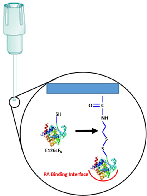
In vivo, proteins are often part of large macromolecular complexes where binding specificity and dynamics ultimately dictate functional outputs. In this work, the pre-endosomal anthrax toxin is assembled and transitioned into the endosomal complex. First, the N-terminal domain of a cysteine mutant lethal factor (LFN) is attached to a biolayer interferometry (BLI) biosensor through disulfide coupling in an optimal orientation, allowing protective antigen (PA) prepore to bind (Kd 1 nM). The optimally oriented LFN-PAprepore complex then binds to soluble capillary morphogenic gene-2 (CMG2) cell surface receptor (Kd 170 pM), resulting in a representative anthrax pre-endosomal complex, stable at pH 7.5. This assembled complex is then subjected to acidification (pH 5.0) representative of the late endosome environment to transition the PAprepore into the membrane inserted pore state. This PApore state results in a weakened binding between the CMG2 receptor and the LFN-PApore and a substantial dissociation of CMG2 from the transition pore. The thio-attachment of LFN to the biosensor surface is easily reversed by dithiothreitol. Reduction on the BLI biosensor surface releases the LFN-PAprepore-CMG2 ternary complex or the acid transitioned LFN-PApore complexes into microliter volumes. Released complexes are then visualized and identified using electron microscopy and mass spectrometry. These experiments demonstrate how to monitor the kinetic assembly/disassembly of specific protein complexes using label-free BLI methodologies and evaluate the structure and identity of these BLI assembled complexes by electron microscopy and mass spectrometry, respectively, using easy-to-replicate sequential procedures.
Machen, A. J., O'Neil, P. T., Pentelute, B. L., Villar, M. T., Artigues, A., Fisher, M. T. Analyzing Dynamic Protein Complexes Assembled On and Released From Biolayer Interferometry Biosensor Using Mass Spectrometry and Electron Microscopy. J. Vis. Exp. (138), e57902, doi:10.3791/57902 (2018).
17427
wp-singular,portfolio_page-template-default,single,single-portfolio_page,postid-17427,wp-theme-bridge,bridge-core-3.0.1,qode-page-transition-enabled,ajax_fade,page_not_loaded,,paspartu_enabled,paspartu_on_top_fixed,paspartu_on_bottom_fixed,qode_grid_1200,qode_popup_menu_push_text_top,qode-theme-ver-28.7,qode-theme-bridge,disabled_footer_top,wpb-js-composer js-comp-ver-6.8.0,vc_responsive
Analyzing Dynamic Protein Complexes Assembled On and Released From Biolayer Interferometry Biosensor Using Mass Spectrometry and Electron Microscopy
Analyzing Dynamic Protein Complexes Assembled On and Released From Biolayer Interferometry Biosensor Using Mass Spectrometry and Electron Microscopy
J. Vis. Exp. (138), e57902, doi:10.3791/57902 (2018).
Machen, A. J., O'Neil, P. T., Pentelute, B. L., Villar, M. T., Artigues, A., Fisher, M. T.
Abstract
In vivo, proteins are often part of large macromolecular complexes where binding specificity and dynamics ultimately dictate functional outputs. In this work, the pre-endosomal anthrax toxin is assembled and transitioned into the endosomal complex. First, the N-terminal domain of a cysteine mutant lethal factor (LFN) is attached to a biolayer interferometry (BLI) biosensor through disulfide coupling in an optimal orientation, allowing protective antigen (PA) prepore to bind (Kd 1 nM). The optimally oriented LFN-PAprepore complex then binds to soluble capillary morphogenic gene-2 (CMG2) cell surface receptor (Kd 170 pM), resulting in a representative anthrax pre-endosomal complex, stable at pH 7.5. This assembled complex is then subjected to acidification (pH 5.0) representative of the late endosome environment to transition the PAprepore into the membrane inserted pore state. This PApore state results in a weakened binding between the CMG2 receptor and the LFN-PApore and a substantial dissociation of CMG2 from the transition pore. The thio-attachment of LFN to the biosensor surface is easily reversed by dithiothreitol. Reduction on the BLI biosensor surface releases the LFN-PAprepore-CMG2 ternary complex or the acid transitioned LFN-PApore complexes into microliter volumes. Released complexes are then visualized and identified using electron microscopy and mass spectrometry. These experiments demonstrate how to monitor the kinetic assembly/disassembly of specific protein complexes using label-free BLI methodologies and evaluate the structure and identity of these BLI assembled complexes by electron microscopy and mass spectrometry, respectively, using easy-to-replicate sequential procedures.
Category
2018, Publications